Intraoral Dental Imaging In Santa Paula

Make A Quick Appointment

santa paula dentist

Intraoral Dental Imaging In Santa Paula

Make A Quick Appointment

Santa Paula Dentist

Welcome to Channel Islands Family Dental

Frequently Asked Questions

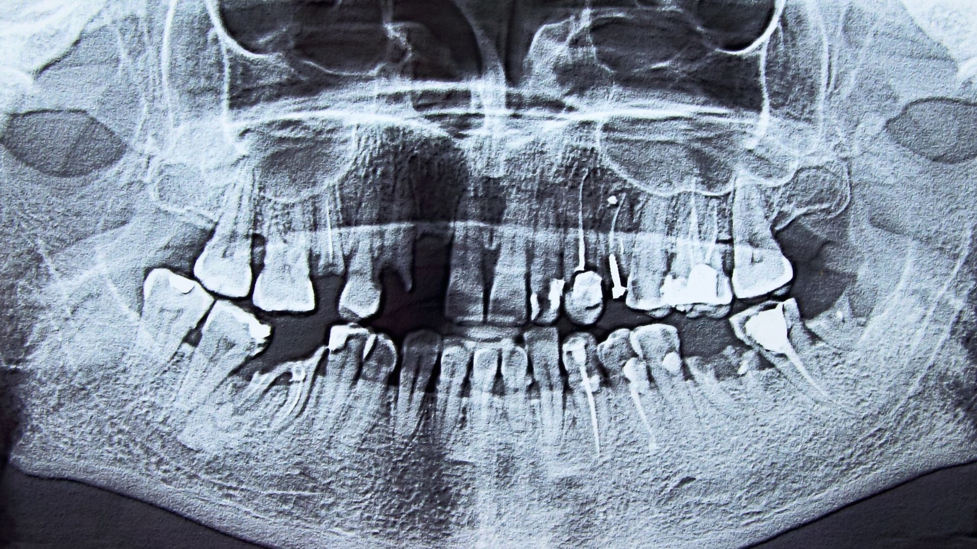
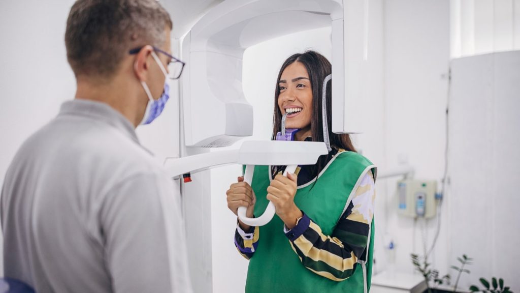
What is intraoral dental imaging?

Intraoral dental imaging involves the use of small cameras or X-ray sensors placed inside the mouth to capture detailed images of the teeth, gums, and other oral structures.

Why is intraoral imaging used in dentistry?

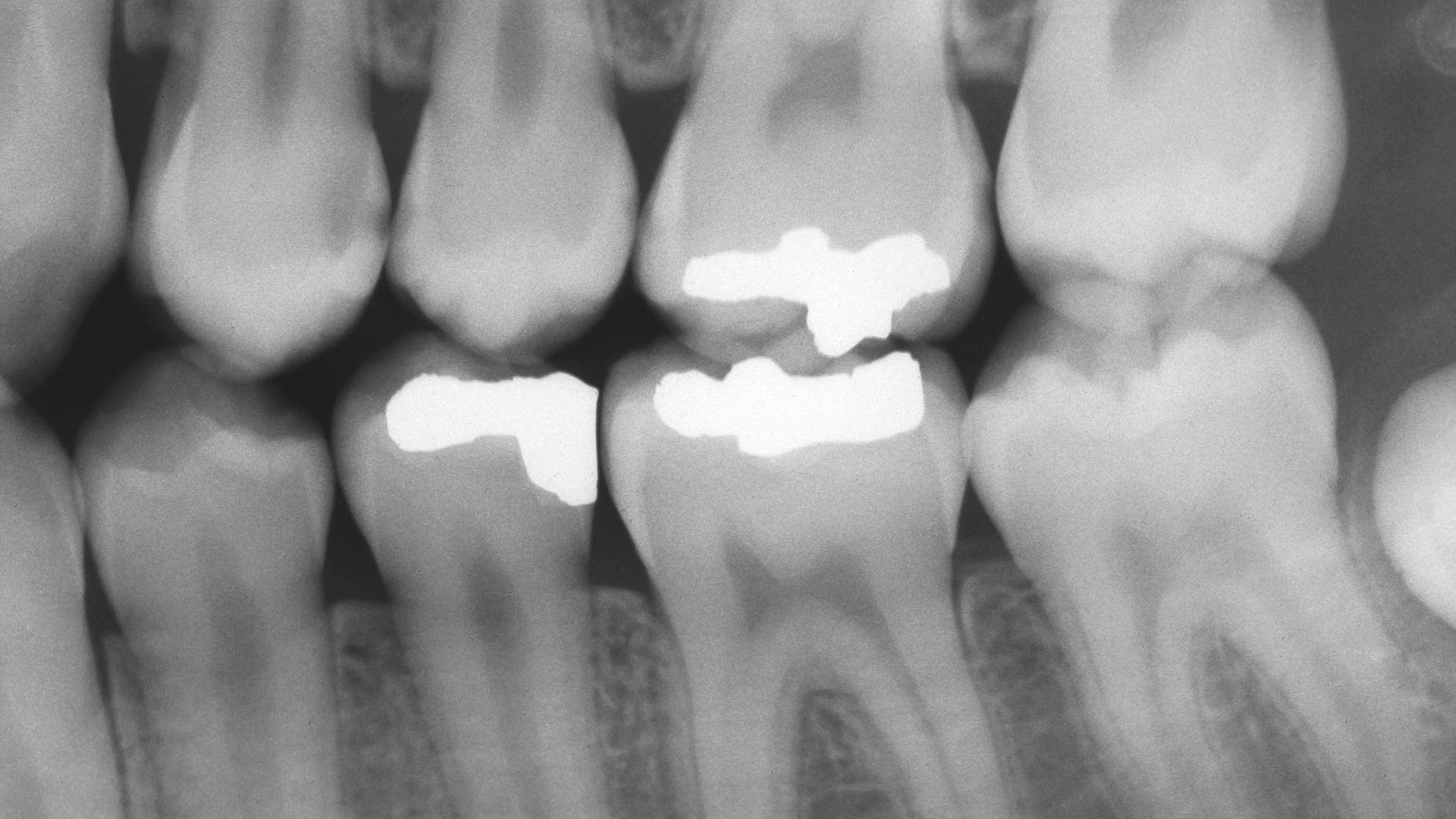
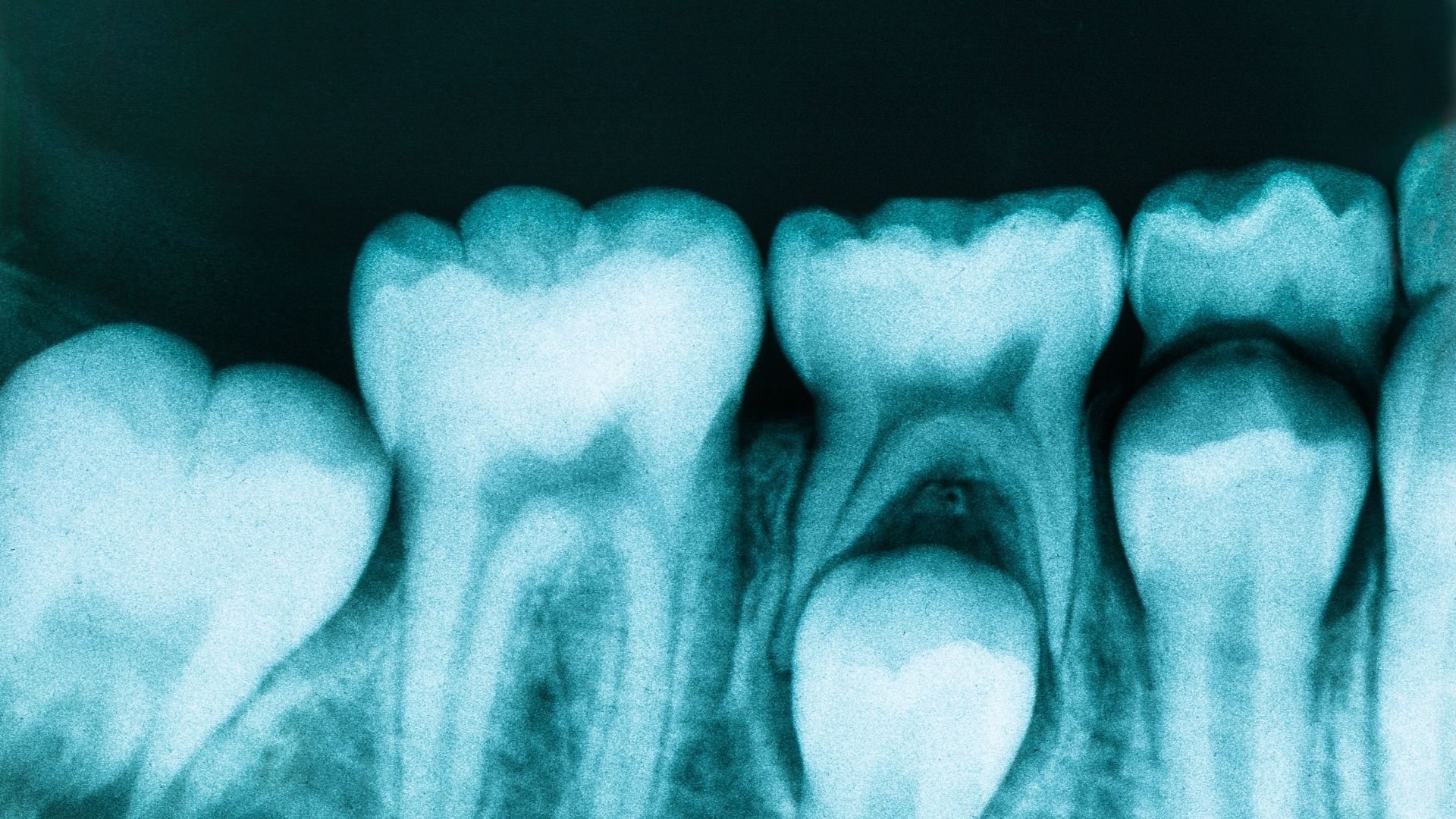
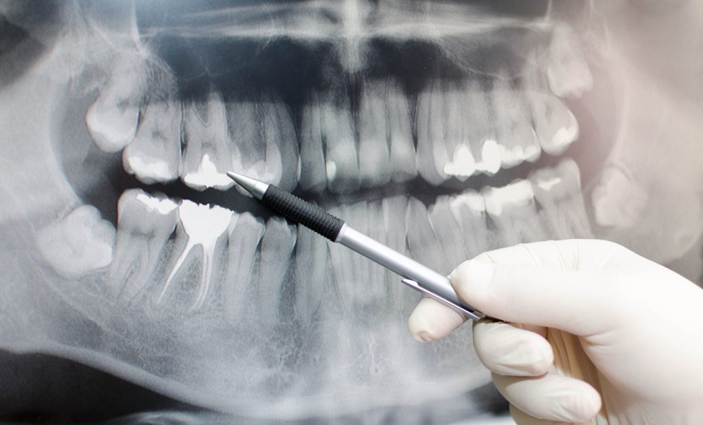
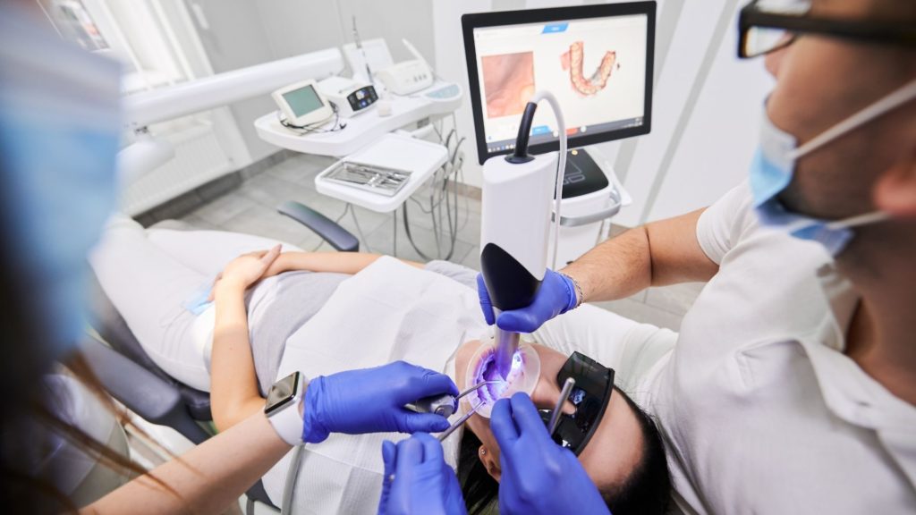
Intraoral imaging provides dentists with detailed views of the oral cavity, aiding in the diagnosis of dental issues such as cavities, gum disease, and structural abnormalities.

Is intraoral imaging safe?

Yes, intraoral imaging is generally safe. It uses low levels of radiation for X-rays, and the equipment is designed to minimize exposure. It is a valuable diagnostic tool with proven safety records.

How often are intraoral images taken during a dental visit?

The frequency of intraoral imaging varies, but it is typically part of routine dental check-ups. Dentists may recommend it more frequently based on individual oral health needs and concerns.

Intraoral Dental Imaging

intraoral dental
Whether for aesthetic or other reasons, it is increasingly common to want to have a perfect, white, and bright smile. All this can be achieved thanks to the advances in technology in dentistry, including Intraoral Dental Imaging in Santa Paula. Now there are several procedures that help improve our smiles. Currently, there are two very common and widely-used procedures to lighten the teeth; both must be performed in the office by dentists. The two treatments act in a similar way, but some aspects make them different.

Intraoral Dental Imaging In Santa Paula

Intraoral Radiographs

The vast majority of dental images are created from the application of X-ray technology; to these devices, we can obtain the necessary information to determine a timely and accurate diagnosis. Intraoral radiographs use radiographic films placed inside the oral cavity. There are different types of radiographs, among them we have the following:

Periapical Radiography

intraoral dental

This type of radiography is one of the most requested. It is also known as dental radiography because thanks to it, you can observe the entire tooth from the tip of the root to the crown, which is the visible part in the mouth. From this radiographic image,you can evaluate the state of the tooth and its adjacent structures such as the bone around it and the supporting tissues. It is useful to determine the progress of tooth decay, the presence of pulp involvement, the presence and progress of any infection and to rule out any fracture or periodontal disease.

Bitewing Or Interproximal Radiographs

intraoral dental imaging

These are radiographs of the same size as the periapical type used with a different technique to obtain the intraoral images that will help us obtain information about a greater number of teeth, but only of the coronary portion of both dental arches; that is to say, of the upper and lower jaw. One of the main reasons for requesting this type of radiography is to determine the presence of dental caries between teeth, which is often difficult to diagnose in a clinical examination. It is also useful for the evaluation of the presence and progression of disease of the supporting structures (periodontium).

Occlusal Radiography

intraoral dental imaging

These radiographs are slightly larger compared to periapical radiographs. In order to obtain this intraoral image, a technique is used in which the patient bites the radiographic plate. With the radiograph, it is possible to evaluate the location of  teeth that have not yet erupted in the mouth or those  in a different position than normal.

intraoral dental imaging

Extraoral Radiographs

Intraoral radiographs are not the only way to obtain dental images. There are also extraoral radiographs. Itt is already very common to obtain images through the use of cone beam computed tomography (CBTC); this type of image generates more information because the image is in different planes as a three-dimensional image (3D). An investigation published in the journal of oral and maxillofacial surgery, conducted by the academic and professional community of WHO determined that the use of cone beam tomography is safe since it ensures safety parameters for the patient in terms of radiation exposure.

The decision of what type of image to request depends on different factors but fundamentally is based on what is to be observed: the position of a tooth, the presence of periodontal disease, fractures, infections, and the evaluation of any treatment, among others. It is important to emphasize that dental x-ray examinations are safe according to the American Dental Association (ADA) since they have a very low level of radiation exposure, which makes the risk of possible harmful effects very small. When considering Santa Paula dental care, trust in our expertise for your dental imaging needs.

More easier to schedule your appointments

Dental X-Rays - Radiografías Dentales

Intraoral Dental Imaging

Intraoral Dental Imaging. Technology in the dental field has been evolving over time, helping both the dentist and the patients obtain better diagnoses in less time and within everyone’s reach.

Intraoral Scanning

This type of intraoral imaging allows us to create a 3D digital file of the patient’s mouth and thus be able to elaborate the work digitally, reducing processing time and often affording greater accuracy. This system will replace the traditional models made from the patient’s mouth, giving greater comfort to the patient.
intraoral dental

Intraoral Dental Imaging In Santa Paula

Financing Options

Payment plans as low as 99/month (*on approved credit)

dental insurance block
care credit
ALPHAEON CREDIT

Our Location

We have the experience and expertise to deliver high quality dental specialty care. Contact us today with your dental questions or to schedule your appointment.

 
Office Hours
Monday – Friday9:00am – 5:00pm
 
Phone Number